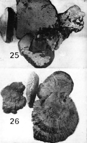
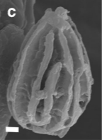
25:菌孔面; 26:菌盖表面。来源:Some tropical species of Ganoderma (Polyporaceae) with pale context担孢子的扫描电镜(SEM)图结果。来源:Taxonomy and phylogeny of polypores with ganodermatoid basidiospores (Ganodermataceae)脉孢新热带灵芝Neoganoderma neurosporum (J.S. Furtado) B.K. Cui & Y.F. Sun 2022 异名: Ganoderma neurosporum J.S. Furtado 1967 Haddowia neurospora (J.S. Furtado) Teixeira 1992
子实体一年生,具侧生柄或无柄,木质。菌盖单生,平展至凸镜形。菌盖表面红褐色至暗褐色,不具漆状光泽,光滑,具同心环状沟纹;菌盖边缘钝。菌肉苍白色至奶油色,软木栓质。菌柄暗褐色,基部略呈空心。菌孔面奶油色至浅肉桂褐色;管壁厚,全缘;菌孔圆形;菌管浅灰褐色。 分布:分布于巴拿马、哥斯达黎加、巴西等新热带地区。中国目前未有分布 讨论:本种具灵芝状担子果和类鸡冠孢芝的担孢子,曾被置于灵芝属和鸡冠孢芝属中,但因其略具平截的担孢子以及拥有特殊的内孢壁纹饰(内孢壁纹饰具担孢子等长的纵向脊突但不具明显的横向脊突)与灵芝属物种和鸡冠孢芝属物种区分开来. 相似物种:与灵芝属物种和鸡冠孢芝属物种较为相似,但本种担孢子略具平截以及拥有特殊的内孢壁纹饰(内孢壁纹饰具担孢子等长的纵向脊突但不具明显的横向脊突)。 新组合描述:
Description: Basidiomata annual, laterally stipitate or sessile, corky. Pilei solitary, flat to convex, up to 15 cm diam and 3 cm thick. Pileal surface reddish brown to dark brown, dull, glabrous, with concentric furrows; margin obtuse, entire. Pore surface cream to pale cinnamon brown; pores circular, 4–5 per mm. Context pallid white or cream, soft corky, up to 2.5 cm thick. Tubes pale greyish brown, up to 1.5 cm long. Stipe dark brown, slightly swollen at base, up to 10 cm long and 1.5 cm diam. Hyphal system dimitic; generative hyphae with clamp connections, colourless, thin-walled, branched, 2–5 μm diam; skeletal hyphae terminal arboriform or unbranched, colourless to pale yellow, 3–7 μm diam. Pileipellis composed of irregularly to slight anticlinal skeletal hyphae and thin generative hyphae. Basidiospores ellipsoid, slightly truncated, pale yellow, IKI–, double-walled with thick walls, endospore wall with longitudinal ridges which equal in length to basidiospores, 16–20 × 11–15 μm.
Notes: The brief description of Neoganoderma neurosporum was taken from Ryvarden (2004b) and Costa-Rezende et al. (2020b). According to the records, N. neurosporum is known from dead wood of deciduous trees in Neotropics. Neoganoderma neurosporum was placed in Haddowia by its similar endospore wall ornamentation, but no obvious traverse ridges were observed in N. neurosporum under SEM. Besides, N. neurosporum formed an independent lineage in the phylogenetic analyses (Fig. 1). More detailed description of N. neurosporum need to be made from future collections.