Leucoagaricus purpurascens T. Guo & Z. W. Ge, sp. nov. FIGURES. 2–3 MycoBank:—MB 845096
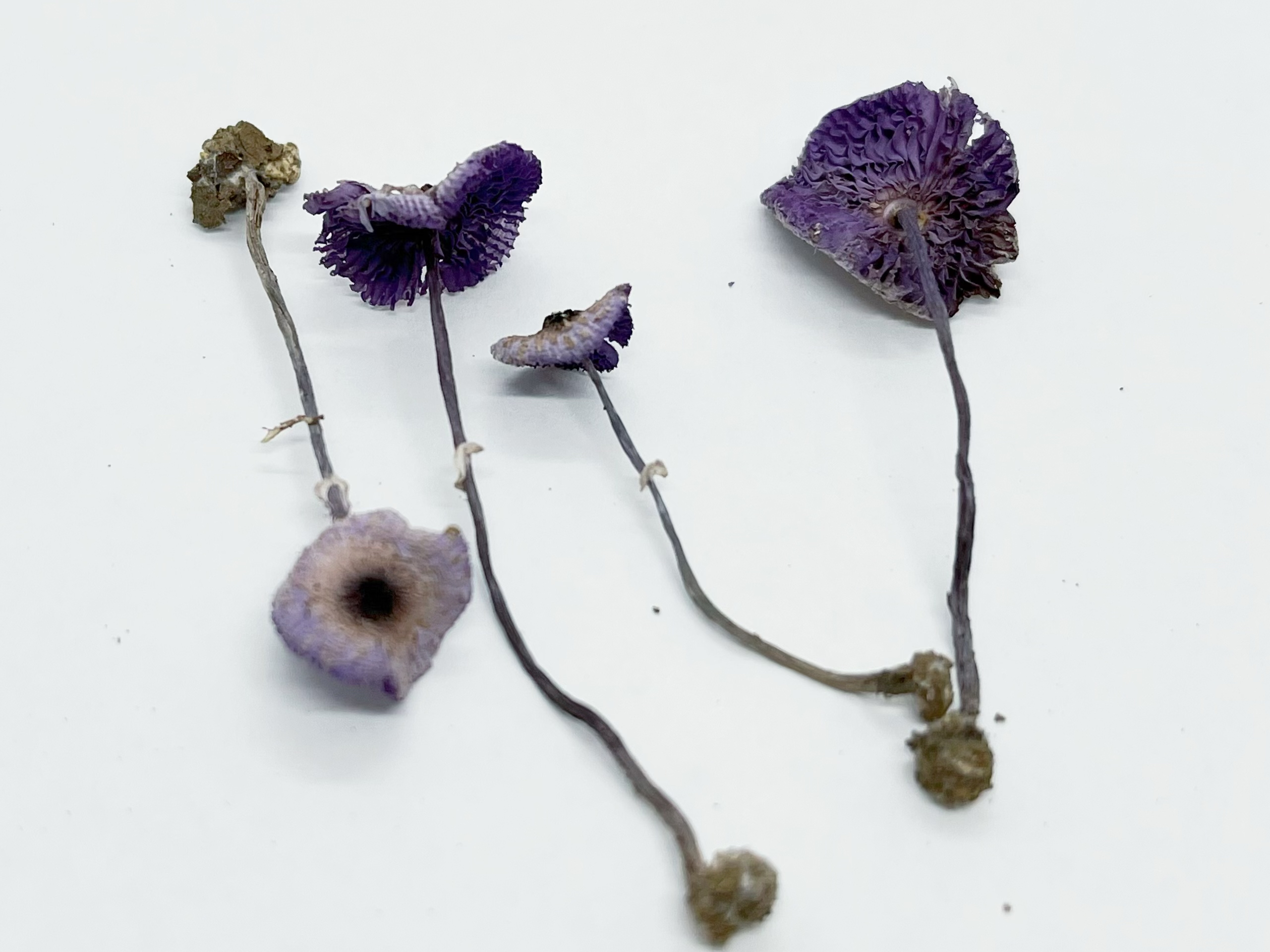
Etymology:—“purpurascens” means becoming purple, referring to the basidiomata turning purple upon drying.
Diagnosis: Similar to Leucoagaricus viriditinctus but differs in the purple changes of basidiomata when dried, larger basidiospores, and the absence of cheilocystidia.
Holotype:—CHINA. Shanghai: Sheshan National Forest Park, on the ground in an evergreen broad-leaved forest, elev. 40 m, 22 September 2020, T. Guo 2137 (HKAS 123023!). GenBank: ITS = OM987458; LSU = OM987455.
Description:
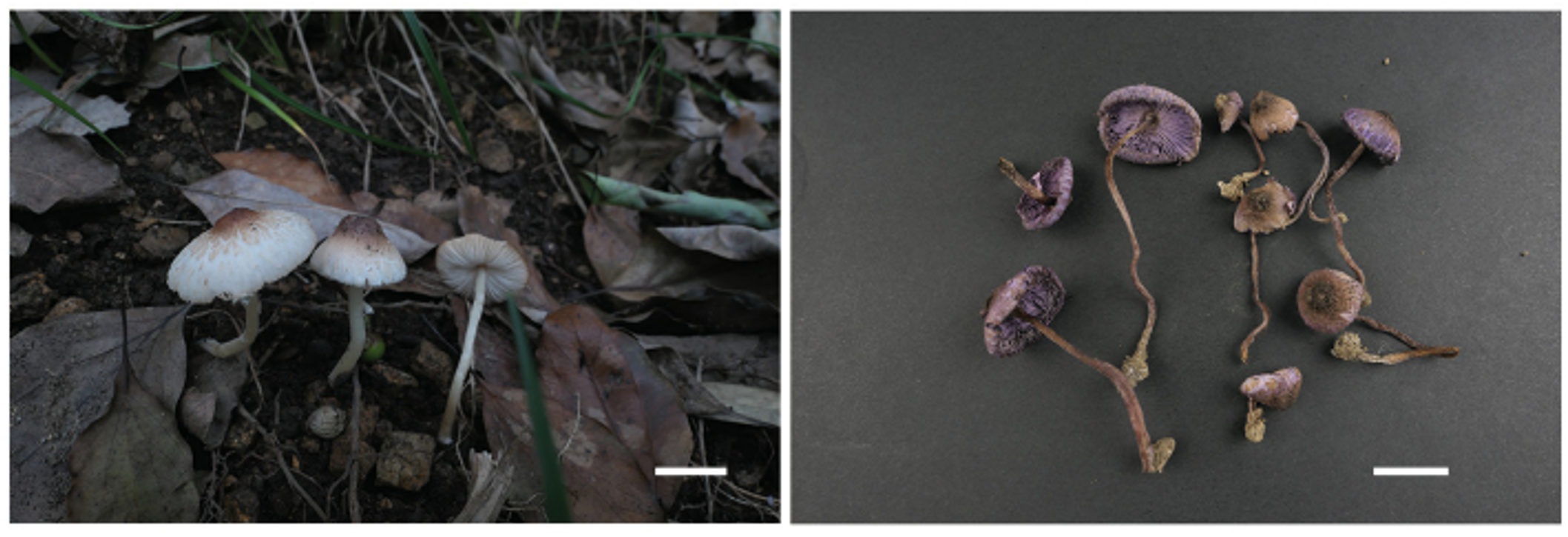
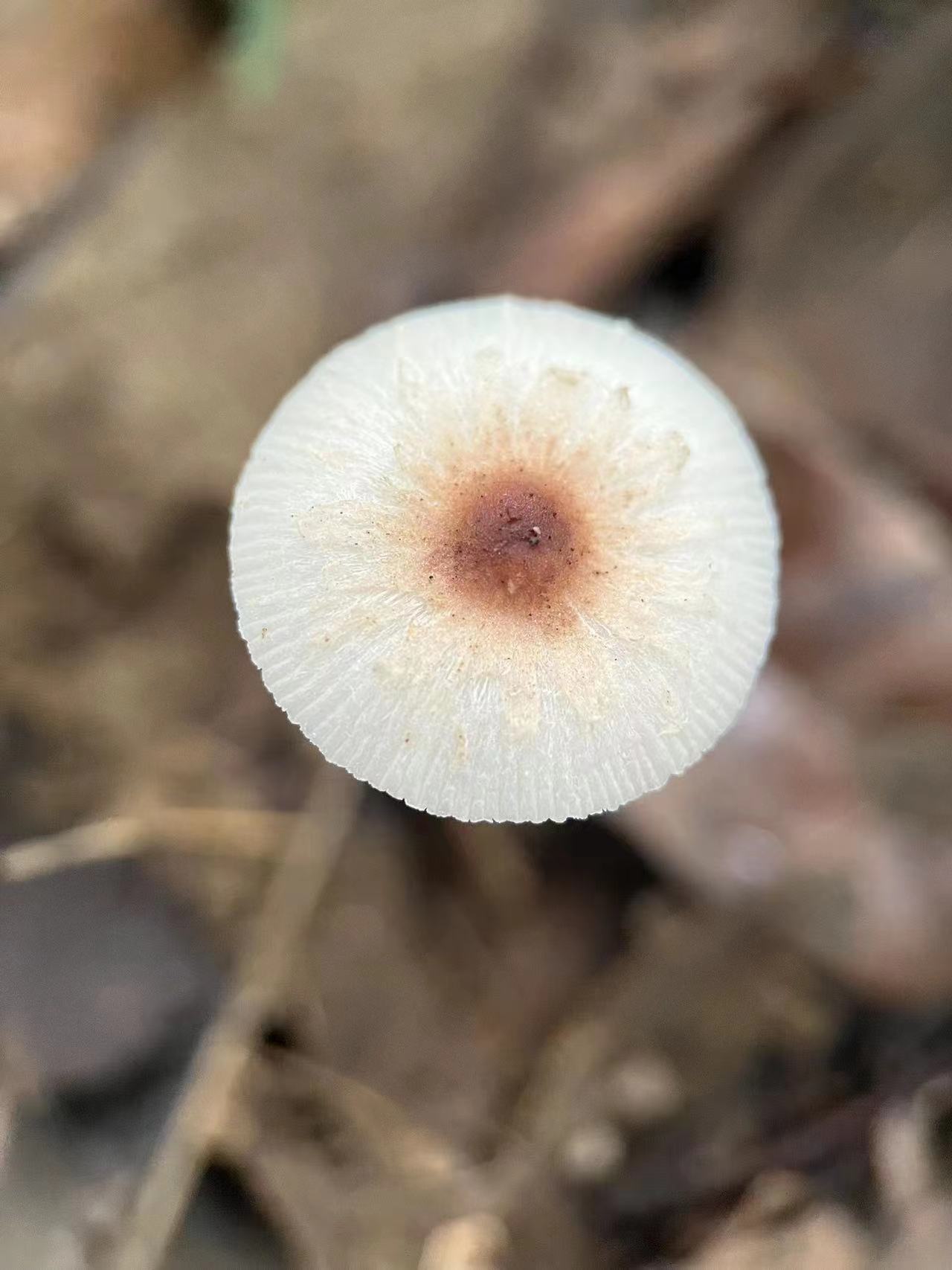
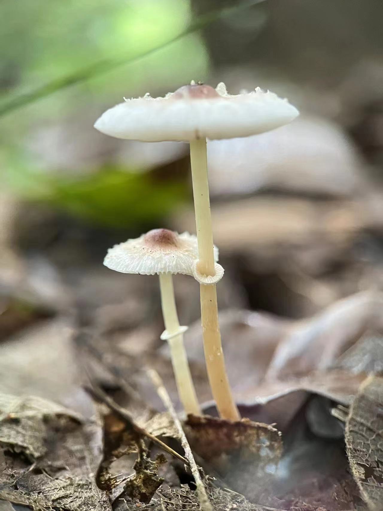
—Basidiomata (FIGURE 2). Pileus 1–2.5 cm diam, when young campanulate, expanding to plano convex to applanate, umbonate; center light brownish (7C3) to dark brown-red (8E4), elsewhere with light brownish squamules on the whitish (9A1) background; Context thin, whitish (9A1); discolours purplish (16B2) to purple (16A4) on drying. Lamellae free, whitish to creamy (6A2), crowded, with lamellulae in 1–2 tiers, edge even; discolours purplish (16B2) to purple (16A4) on drying. Stipe 2.5–5 × 0.1–0.2 cm, whitish (9A1), slightly attenuate upwards, sometimes slightly curved at the base, smooth, hollow, not brittle; stipe context white; Annulus present, sometimes disappearing, with light brownish (6C5) superior edge, membranous. Odor none. Taste not recorded.
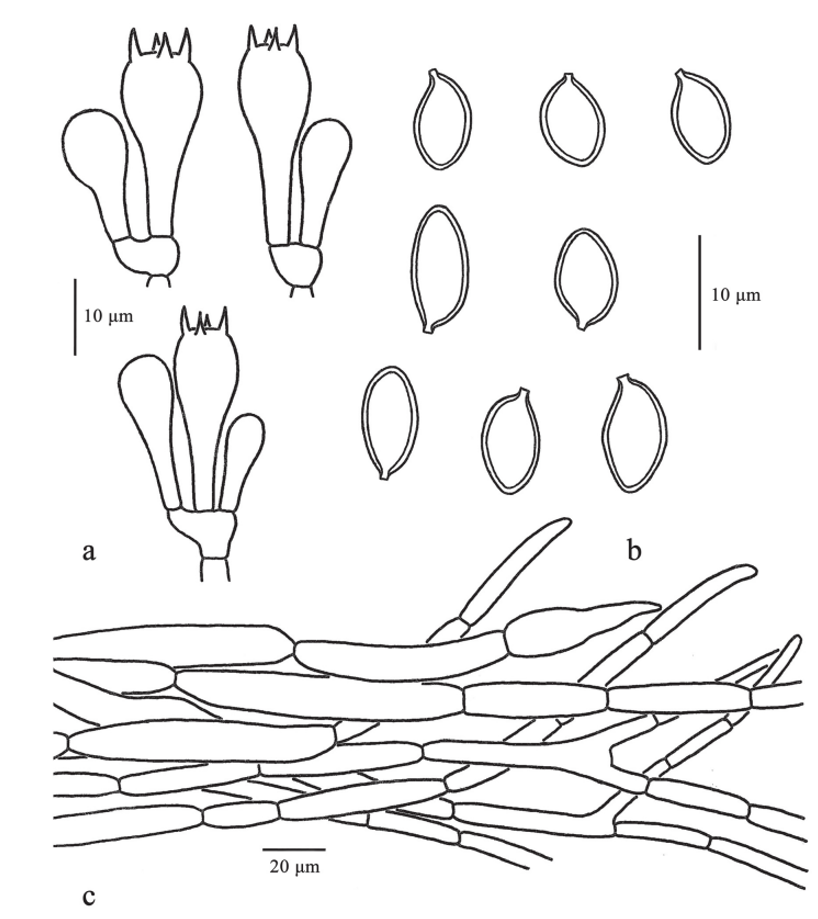
Basidiospores (FIGURE 3) [71/3/3] 8.0–10.0 (10.5) × (4.0) 4.5–6.0 μm (mean 9.2 ± 0.7 × 5.1 ± 0.5 μm), Q = (1.5) 1.6–2.0, Qav = 1.79 ± 0.15, amygdaliform, (sometimes ellipsoidal to oblong) in side view, ovoid in front view, hyaline, smooth, slightly thick-walled (about 0.5 μm), dextrinoid, without germ pore, metachromatic in Cresyl Blue. Basidia (24.0) 24.5–31.0 (32.0) × (9.0) 9.5–10.5 (11.0) μm, clavate, four-spored; sterigmata up to 3 μm. Cheilocystidia and pleurocystidia not observed. Lamella trama subregular, made up of slightly inflated subcylindrical hyphae, hyaline, thin-walled. Pileus covering a cutis with radially arranged and sometimes branched repent hyphae, subcylindrical, 7.5–12.5 μm diam, occasionally with brownish yellow pigments on the hyphal wall and in vacuoles. Stipe covering a cutis made up of cylindrical hyphae and elements, colourless, (4) 7–10 μm wide. Clamp connections not observed.
Habitat and distribution:—Gregarious or scattered in the subtropical evergreen broad-leaved forest; currently only known from the type locality.
Additional specimens examined:—CHINA. Shanghai City, Sheshan National Forest Park, on the roadside in a subtropical evergreen broad-leaved forest, elev. 35 m, 4 August 2021, T. Guo 2323 (HKAS 123024); same locality, 4 August 2021, T. Guo 2324 (HKAS 123025).
Discussion: The species newly described in this study, La. purpurascens, is characterized by the purple changes of basidiomata when dried, its umbonate pileus covered with light brownish squamules, absence of cheilocystidia, and the relatively larger basidiospores (FIGURES 2–3). Phylogenetic evidence also suggests that La. purpurascens differs from its close relatives (FIGURE 1).
Cheilocystidia are typically present in species of Leucoagaricus. However, it is reported that broadly clavate to pyriform cheilocystidia were observed in some collections of La. vinditinctus, although no cheilocystidia were observed from its type specimen (Liang et al. 2010). In the present study, repeated observations of the lamella edges of La. purpurascens collections only found narrowly clavate to clavate cells (19.0–27.0 × 6.0–8.5 [10.0] μm) intermixed with mature basidia. These narrowly clavate to clavate cells are smaller than basidia (24.5–31.0 × 9.5–10.5 μm), suggesting these narrowly clavate to clavate cells are young basidia instead of cheilocystidia and indicating the absence of cheilocystidia of this species.
In the phylogram (FIGURE 1), La. purpurascens is sister to La. viriditinctus. These two jointly form a sister clade with La. virens Y. R. Ma, Z. W. Ge & T. Z. Liu. However, La. viriditinctus differs from La. purpurascens by presenting dark blue changes of basidiomata when bruised or dried, smaller basidiospores (7.0–8.5 × 4.0–5.0 μm), and having broadly clavate to pyriform cheilocystidia and mainly from tropical regions (Liang et al. 2010; Retnowati 2015), while La. virens, described from a temperate region, differs from La. purpurascens in having clavate to broadly clavate cheilocystida, and presenting dark green to light green reaction when the basidiomata were bruised or dried (Ma et al. 2022).
Morphologically, La. purpurascens also resembles La. atrosquamulosus (Hongo) Z. W. Ge & Zhu L. Yang (Yang & Ge 2017). However, La. atrosquamulosus can be distinguished from La. purpurascens by its larger basidiomata, blackish brown squamules on the pileus, the presence of cheilocystida and smaller basidiospores (5–7 × 3.5–4 μm). Several other Leucoagaricus and Leucocoprinus species with bruising reactions exist in China, viz. La. atroazureus, La. flavovirens J. F. Liang, Zhu L. Yang & J. Xu, La. virens Y. R. Ma, Z. W. Ge & T. Z. Liu, and Lc. viridiflavus (Petch) E. Ludw. However, these species display blue or green reactions and have cheilocystidia (Ge et al. 2019; Ma et al. 2022). Leucoagaricus purpureoruber also stains purple on drying, but it has larger basidiomata, clavate to fusiform cheilocystidia and basidiospores with an apical germ pore (Yang & Ge 2017).