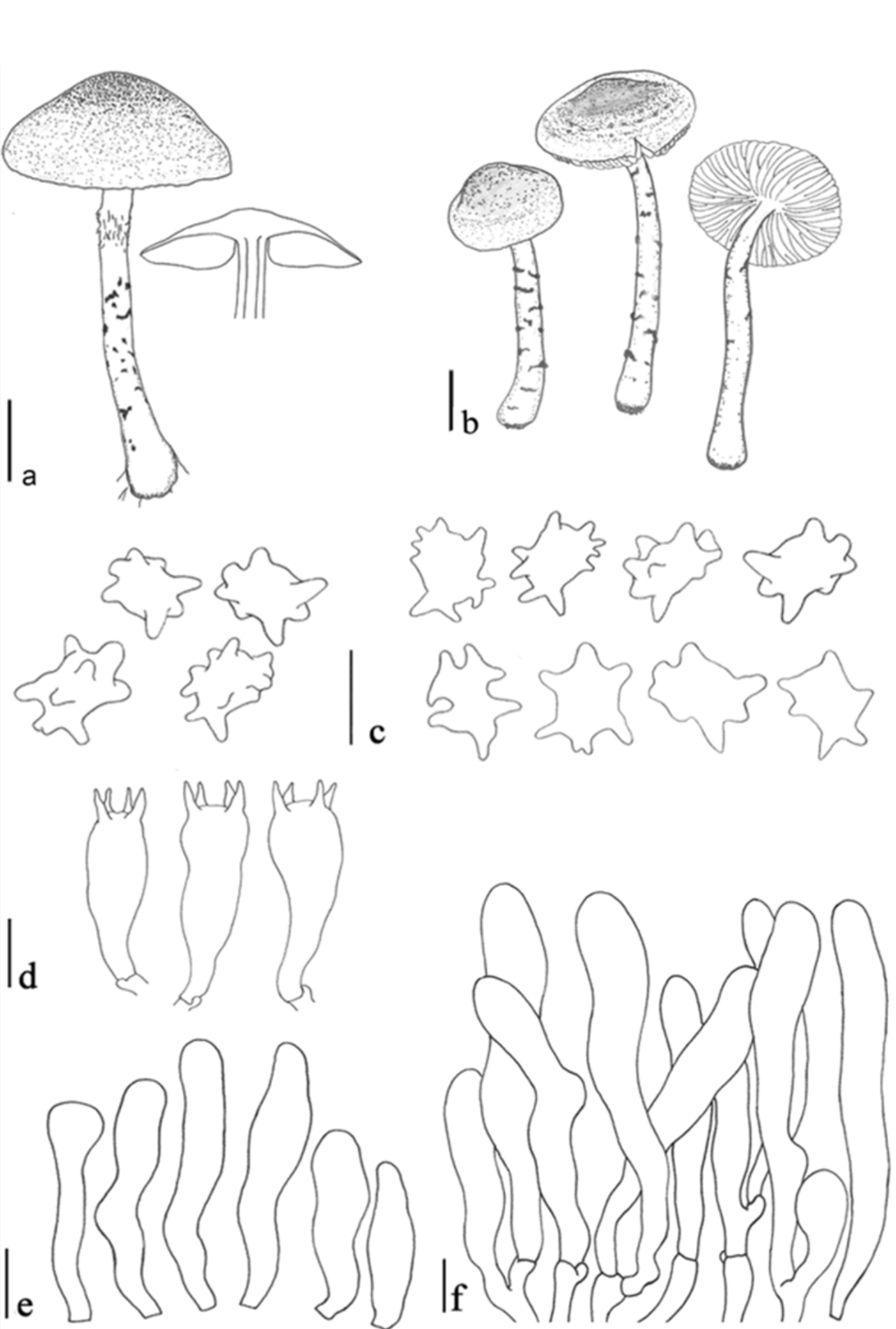
Verrucospora vulgaris Pegler, Kew Bull.,Addit. Ser. 6: 384 (1977) (Figure 1 and 2)
≡ Lepiota verrucispora Beeli, Bull. Soc. R.Bot. Belg. 64: 218 (1932).
≡ Verrucospora verrucispora (Beeli) E.Horak, Ber. schweiz. bot. Ges. 77: 363(1967), nom. inadmiss.
Misapplied names -Verrucospora flavofusca(Henn.) J lich, Biblthca mycol. 85: 401.1982; Horakia flavofusca (Henn.)Oberw., Sydowia 28: 359 (1975)
Pileus 15-30 mm, first parabolic,expanding to subumbonate, often convexto plano-convex, with straight margin, withcompletely yellowish brown (5F6-7)surface, often with darker colour, greenish-brown (5D3), then surface breaking,yellowish brown (5F6-7) at centre or umbo,with fine squamules around center, withfine squamules more distant from eachother around umbo toward margin, whendry squamules turning dark brown to blackon greyish yellow (2B4) background, atmargin with velar squamules or finesquamules; margin fringed when matureand exceeding lamellae. Lamellae not reallyfree, but sinuate, slightly crowded, with 3series of lamellulae, ventricose to broadlyventricose 4-6 mm wide, pastel yellow tolight yellow (2A4, 2A5), with eroded andconcolorous edge, sometimes turningslightly bluish white (20A2) at edge. Stipe40-66 × 2.8-5 mm, cylindrical, some timewider at base or with bulb-like, 5-6 mmwide base, yellowish with (2A4) backgroundat apex to base zone, slightly fibrillose andconcolorous with background at apex,sometimes grayish-yellow (4B3, 4B4) frommiddle zone to base, glabrous or squamuloseat annular zone downward base, conclorouswith background, turned light brown(6D5-6) to dark brown (7F4, 7F5) andblack when dried, with white to yellowish-white (4A2) rhizomorphic mycelium atbase and connected to substrate. Annuluswith annular zone of yellowish-with (2A4)fibrillose, squamulose, grayish-yellow tolight brown (4B3, 6D5-6). Context lightyellow to grayish-yellow (2A5, 2B4) and4-4.5 mm wide in pileus, sometimes turnedbluish-white (20A2) but not much;in stipe, concolorous with surface,hollow, with white fibrils in hollow.Odour similar to odour of Leucoagaricusnaucinus. Taste unknown. Spore printwhite in all collections.
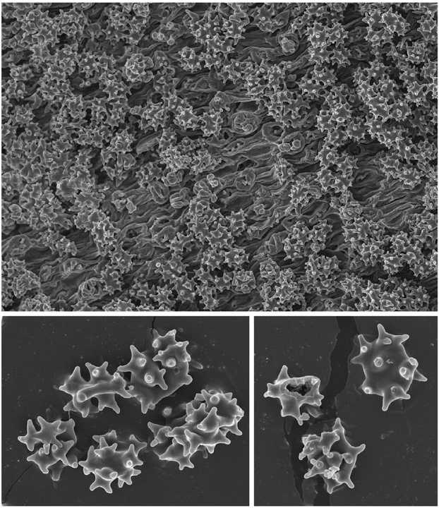
Basidiospores [100, 5, 5] 6.4-8.6 × 3.6-5.8 μ m, avl × avw = 7.3 x 5.0 μ m, Q =1.25-1.63, avQ = 1.48, in side view slightlyangular or verrucose in young stage,developing numerous papillae, becomingirregularly angular, pentagonal or star-like,with irregular angles in frontal view,dextrinoid, congophilous, cyanophilous,not observed in Cresyl Blue. Basidia 26-28× 7.8-8.4 μ m, clavate, slightly curved atbase, thick-walled, hyaline, 4-spored, rarely1- or 2-spored. Lamella edge sterile, withcrowded cheilocystidia. Cheilocystidia 23-45 × 4.9-6.5 μ m, cylindrical, or slightlyirregularly cylindrical, sometime cylindricaland swollen at apex, rarely short narrowlyclavate to slightly utriform, slightly thick-walled, colourless. Pleurocystidia absent.Pileus covering a trichoderm made up oferect, irregular, narrowly clavate, narrowlycylindrical elements, 60-88 × 13.6-15.2 μ m,brown-walled, with brown parietal andintracellular pigment, often with shortclavate elements under long elements, 25-35 x 9-12 μ m, with pale brown parietal andintracellular pigment. Squamules on stipecovering with same structure as those ofpileus covering. Clamp connections presentbut not abundant.
Habitat and distributions: Growingsolitary or in small groups, on humus soilwith high nutrient source. Found in 2kinds of deciduous forest; Huai Kok Ma(at Chiang Mai Province) is a forest withdominance with Castanopsis spp. andLithocarpus echinops with abundant leavecompost in forest floor; and a forest DoiTung (at Chiang Rai Privince) is forestdominant with Pinus kesiya and coffeegarden.
Material examined: THAILAND -Chiang Mai Prov., Muang Dist., SuthepSub-Dist., Huai Kok Ma Village,N18 o 48.62' E098 o 54.60', alt. 1145 m., 28June 2010, J.M. Birkebak P81(MFLU100599); ibidem, 29 June 2010, P.Sysouphanthong P83(MFLU10 0601);ibidem, 29 June 2010, P. SysouphanthongP86 (MFLU10 0604); ibidem, 10 July 2010,J.M. Birkebak P109 (MFLU10 0627);Chiang Rai Prov., Mae Fah Luang Dist.,Doi Tung Sub-Dist., N20 o 17.37', E99 o49.08', Alt. 860 m., 17 July 2010, P.Sysouphanthong P122 (MFLU10 0640).
疣孢环柄菇 图112
Verrucospora vulgaris Pegler. Kew Bull. Add. Ser.6:384.1977.
Lepiota verrucospora Beeli. Bull. Soc. Roy. Bot. Belg.64:218. fig.23.1932.
Verrucospora verrucospora(Beeli) Horak. Ber. Schweiz. Bot. Ges.77:363. fig.1,1967(nom. inadmiss.).
担子果小型。菌盖直径2~3cm,初半球形,后扁平至平展,黄色至绿黄色,中部橄榄褐色至灰橄榄褐色,菌盖表面密被淡褐色至淡灰色细小鳞片;菌肉淡黄色,较薄,伤后有蓝色色调;菌褶离生,柠檬黄色至硫磺色(干后变为深褐色至近黑色),较密,不等长,边缘腹鼓状,最宽达0.5cm。菌柄长5~6.5cm,直径0.3~0.5cm,近圆柱形或向上稍变细;表面白色至淡黄色,被淡褐色至淡灰色丝状鳞片;中空;菌环易消失。
担子28~35×8~11μm,棒状,多具4孢梗。担孢子9~11×6.5~9μm(含疣刺)或6~8×4~5μm(不含疣刺)[Q=(1.06)1.22~1.57(1.75),Q=1.40±0.12],椭圆形至宽椭圆形,无色透明;表面有明显的疣或刺(长达2μm);壁略厚,类糊精质,刚果红中变红;无芽孔;小尖明显。褶缘囊状体20~35(60)×5~8μm,棒状、窄棒状至近梭形。侧生囊状体阙如。菌盖表面鳞片由呈毛状排列的细胞[25~70×5~10(20)μm]组成,常具淡褐色至淡黄色胞内色素。锁状联合存在于担子果各部位。
生境:夏季生于热带季节性雨林地上。
世界分布:非洲和亚洲的热带地区。
模式产地:喀麦隆。
研究标本:海南:五指山市,五指山,2010年7月31日,葛再伟2557(HKAS 60233)。
讨论:疣孢环柄菇V.vulgaris的主要特征是菌盖或多或少黄色至绿黄色,菌盖表面被淡褐色至淡灰色鳞片,菌褶柠檬黄色至硫磺色,菌柄白色至淡黄色,担孢子表面有明显的疣或刺;锁状联合存在于担子果各部位。
在形态上,V.flavofusca(Henn.)Jülich似乎酷似疣孢环柄菇,但二者是否为同物需要进一步研究。该种原初是置于丝盖伞属的,即Inocybe flavofusca Henn.,据记载其菌褶初为黄色、后变橄榄锈褐色,担孢子橄榄褐色(Hennings,1902)。是否确实如此,不得而知,只有研究模式标本方可解决此疑问。
Sysouphanthong等(2013)曾报道泰国产疣孢环柄菇,本卷作者在泰国清迈附近林中也曾采到此种(凭证标本HKAS 43818)。