返回搜索
Logo Pic

黄鳞囊小伞

Cystolepiota luteosquamulosa


T. Bau and X. Y. Zhou 2024

基本信息 General Info.

  • 通用中文名Chinese name:黄鳞囊小伞
  • 拉丁学名Scientific name:Cystolepiota luteosquamulosa T. Bau and X. Y. Zhou 2024
  • 科Family:疣孢环柄菇科Verrucosporaceae
  • 属Genus:囊小伞属Cystolepiota
  • 分布Distribution:中国东北地区
  • 有分布的省区Provinces:吉林省,黑龙江省(文献记载)
    查看具体分布情况
  • 生态Ecology:单生或散生于混交林的枯叶或土壤上。
  • 营养类型Nutrition Type:腐生Saprotrophic: 分解死亡生物体或有机物获取养分
  • 置信指标Confidence:★★★

物种介绍

DefaultPicture

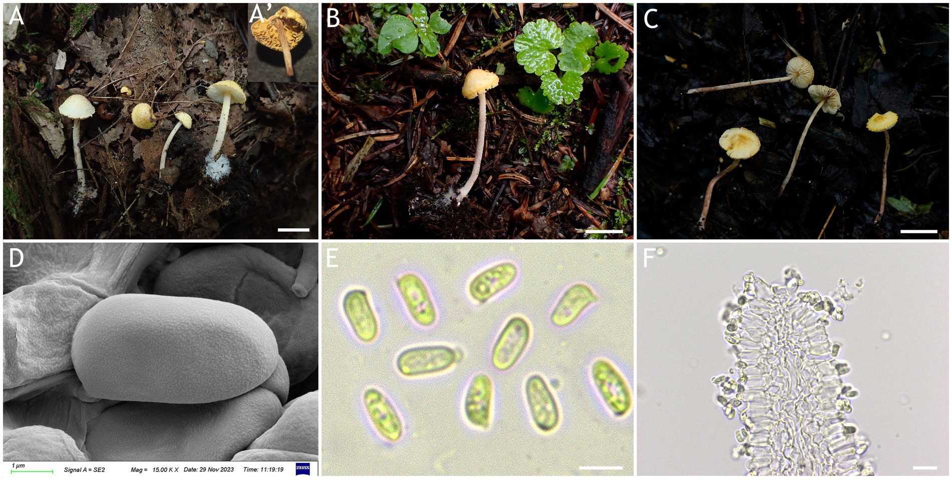
A-C:担子果;A':干标本;D:SEM下担孢子;E:光镜下担孢子;F:子实层末端。A,D-F:HMJAU67711(主模式);B:HMJAU67809;C:HMJAU67810。比例尺:A-C=1cm
来源文献:Four new species of Cystolepiota (Agaricaceae, Agaricales) from northeastern China

黄鳞囊小伞是2024年发现于吉林省的一种囊小伞,其显著特征是整个子实体被有淡黄色至灰黄色的鳞片,无褶缘和侧生囊状体。

形态简述

担子果小。菌盖淡黄色至灰黄色,被灰黄色至黄褐色的疣状至近锥形鳞片,幼年时边缘有菌幕残片。菌褶离生,白色,密集,干后橙褐色。菌柄淡红棕色,有少量白色至淡黄色丛毛状鳞片。担孢子4.9–6.3 (−6.6) × (−2.0) 2.4–3.0 (−3.3) μm,长椭圆形至圆柱形,透明,非淀粉质非类糊精质。无囊状体。

相似物种

南美洲的C. luteohemisphaericum(原隶属于Cystoderma)也拥有黄色菌盖,但其菌盖上有细小的云母状粉末(micaceous-mealy),略有辐射状排列的纹路,担孢子宽椭圆形至椭圆形。C. icterina同样有黄色菌盖,但其菌盖表面被少量丛毛状至粉末状鳞片,担孢子更小(3.5–4.5 × 2.5 μm),类糊精质,有褶缘囊状体。

其他

美国的一份标本(https://www.inaturalist.org/observations/147467243)ITS序列鉴定为本物种。然而,形态学上这份标本和黄鳞囊小伞差异较大(缺乏黄鳞)。

物种描述

文献中物种描述

  • 以下描述摘自相关参考文献,由文字识别而来,请以原文为准。
  • 部分文献可能存在描述过时、描述错误或者描述片面等情况,仅供参考。
  • 外文描述的参考翻译系网站管理者的个人翻译,详请参见原文。

描述 1 来源:Four new species of Cystolepiota (Agaricaceae, Agaricales) from northeastern China
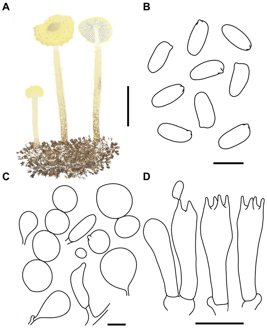
类型:原白(物种发表时的原始描述)

3.2.3 Cystolepiota luteosquamulosa T. Bau and X. Y. Zhou, sp. nov.
MycoBank number: MB 849380 (Figures 7, 8).
Diagnosis: C. luteosquamulosa is distinguished from other Cystolepiota species by its light yellow to greyish yellow pileus, with greyish yellow to dark yellow warty to subpyramidal squamules, light reddish brown stipe with white to light yellow floccose squamules, and pleurocystidia and cheilocystidia absent.
Etymology: “luteo-” means yellow, and “luteosquamulosa” refers to the yellow squamules on the pileus.
Type: China, Jilin province, Jiaohe City, Hongyegu, September 6, 2021, coll. T. Bau and X. Wang (HMJAU67711), Holotype!
Description: Basidiomata small. Pileus 0.8–1.4 cm, hemispherical to obtusely conical when young, expanding to plano-convex or applanate with a slightly umbonate center with age, light yellow (1A4–A8) to greyish yellow (2B5–B8), with greyish yellow (2C7–C8) to dark yellow (3C5–C8) warty to subpyramidal squamules; margin appendiculate with veil remnants when young, and then finely appendiculate, concolorous with pileus; context white, thin. Lamellae free, white to cream, crowded, 0.1–0.3 cm wide, with 1–3 tiers of lamellulae, drying brownish orange (5C2–C6). Stipe 2.5–5.6 × 0.1–0.2 cm, subcylindrical, light reddish brown (7E5–E8), with some floccose squamules, white to light yellow (1A2–A8), with conspicuous white mycelia on the base. Annulus not visible. Odor and taste not recorded (Figures 7A–C, 8A).
Basidiospores [120,4,4] 4.9–6.3 (−6.6) × (−2.0) 2.4–3.0 (−3.3) μm, Q = 1.70–2.63, Qv = 2.10, long ellipsoid to cylindrical, hyaline, smooth-walled under the LM, finely punctate under SEM, inamyloid, non-dextrinoid, metachromatic in cresyl blue, cyanophilous. Basidia 15–23 × 4–7 μm, clavate, 4-spored, sometimes 2-spored. Lamellar trama regular. Pleurocystidia and cheilocystidia absent. Squamules composed of loosely-arranged globose, subglobose, ovoid, 13–62 μm in diam., rarely gourd-shaped or fusiform, 17–42 × 7–16 μm, sometimes 2–4 cells are connected in a string, smooth-walled, slightly thick-walled, hyaline, or orange white (5A2–A3). Clamp connections present in all structures (Figures 7D–F, 8B–D).
Habitat: Solitary or scattered on dead leaves or soil of mixed forest.
Distribution: Northeastern China.
Additional specimens examined: China, Jilin Province, Jiaohe City, Qianjin forest farm, July 24, 2022, coll. T. Bau and L. Y. Zhu (HMJAU67810); Dunhua City, State Forest farm, July 27, 2022, coll. T. Bau, W. N. Hou and F. Guo (HMJAU67808, HMJAU69060); Huadian City, Red Rock National Forest Park, August 28, 2023, coll. T. Bau and H. Cheng (HMJAU67807). Heilongjiang Province, Yichun City, Xing’an National Forest Park, July 25, 2023, coll. T. Bau and W. N. Hou (HMJAU67809).
Notes: Macromorphologically, both C. luteosquamulosa and C. luteohemisphaerica have yellow pileus. But in the latter, the pileus is radially veined and micaceous-mealy, with broadly elliptical to elliptical basidiospores (Saar and Laessoe, 2008). C. icterina also has a yellow pileus, but it is easy to distinguish from C. lutesquamulosa by its pileus surface with finely floccose-farinose squamules, by its smaller (3.5–4.5 × 2.5 μm) and dextrinoid basidiospores and by the presence of cheilocystidia (Knudsen, 1978).

序列信息

如何下载序列?

分子类型 标本号 Version No. 序列 文献 来源地
DNAHMJAU 67711[主模式]OR233619ITS中国 吉林省 Jiaohe City, Hongyegu
DNAHMJAU 67711[主模式]OR240263LSU中国 吉林省 Jiaohe City, Hongyegu
DNAHMJAU 67711[主模式]PP465910rpb2中国 吉林省 Jiaohe City, Hongyegu
DNAHMJAU 67810OR584138ITS中国 吉林省 Jiaohe City, Qianjin forest farm
DNAHMJAU 67810OR584132LSU中国 吉林省 Jiaohe City, Qianjin forest farm
DNAHMJAU 67810PP465914rpb2中国 吉林省 Jiaohe City, Qianjin forest farm
DNAHMJAU 67810PP465899tef1中国 吉林省 Jiaohe City, Qianjin forest farm
DNAHMJAU 67808OR584136ITS中国 吉林省 Dunhua City, State Forest farm
DNAHMJAU 67808OR584130LSU中国 吉林省 Dunhua City, State Forest farm
DNAHMJAU 67808PP465912rpb2中国 吉林省 Dunhua City, State Forest farm
DNAHMJAU 67808PP465900tef1中国 吉林省 Dunhua City, State Forest farm
DNAHMJAU 69060OR936324ITS中国 吉林省 Dunhua City, State Forest farm
DNAHMJAU 67807OR584135ITS中国 吉林省 Huadian City, Red Rock National Forest Park
DNAHMJAU 67807OR584129LSU中国 吉林省 Huadian City, Red Rock National Forest Park
DNAHMJAU 67807PP465911rpb2中国 吉林省 Huadian City, Red Rock National Forest Park
DNAHMJAU 67809OR584137ITS中国 黑龙江省 Yichun City, Xing’an National Forest Park
DNAHMJAU 67809OR584131LSU中国 黑龙江省 Yichun City, Xing’an National Forest Park
DNAHMJAU 67809PP465913rpb2中国 黑龙江省 Yichun City, Xing’an National Forest Park
DNAHMJAU 67809PP465902tef1中国 黑龙江省 Yichun City, Xing’an National Forest Park

涉及该物种的相关文献

  1. Four new species of Cystolepiota (Agaricaceae, Agaricales) from northeastern China

    ,

    2024. Xian-Yan Zhou & Tolgor Bau. Front. Microbiol. 15

        https://doi.org/10.3389/fmicb.2024.1358612