√ 此物种的词条得到了初步审定。
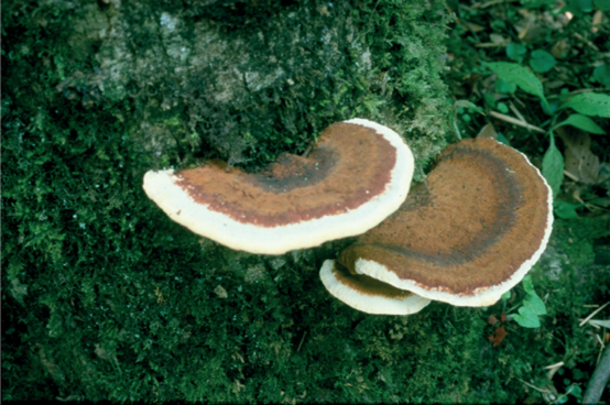
Trachydermella tsunodae
(Yasuda ex Lloyd) B.K. Cui & Y.F. Sun 2022
√ 此物种的词条得到了初步审定。
基本信息 General Info. |
|
类型:完整的描述(摘录自专著、论文等)
序列信息
| 分子类型 | 标本号 | Version No. | 序列 | 文献 | 来源地 |
| DNA | WD2034 | AB588989.1 | ITS | ↗ | 日本 茨城 |
| DNA | WD2034 | AB368069.1 | LSU | ↗ | 日本 茨城 |
| DNA | WD2034 | AB368127.1 | rpb2 | ↗ | 日本 茨城 |
Species diversity, systematic revision and molecular phylogeny of Ganodermataceae (Polyporales, Basidiomycota) with an emphasis on Chinese collections
,2022. Sun Y-F, Xing J-H, He X-L, Wu D-M, Song C-G, Liu S, Vlasák J, Gates G, Gibertoni TB, Cui B-K. Studies in Mycology 101: 287–415
https://doi.org/10.3114/sim.2022.101.05Taxonomy and phylogeny of polypores with ganodermatoid basidiospores (Ganodermataceae)
,2020. D. H. Costa-Rezende, G. L. Robledo, E. R. Drechsler-Santos, M. Glen, G. Gates, B. R. de Madrignac Bonzi et al.. Mycological Progress 19: 725–741
https://doi.org/10.1007/s11557-020-01589-1Taxonomic study on a threatened polypore, Polyporus pseudobetulinus, and a morphologically similar species, P. subvarius
,2011. Sotome K, Hattori T, Ota Y.. Mycoscience 52: 319–326.
https://doi.org/10.1007/S10267-011-0111-X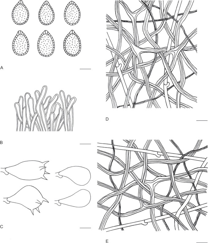
显微特征(绘自Dai 3221c)。A.担孢子 B.表皮顶端细胞 C.担子和幼担子 D.菌髓菌丝 E.菌肉菌丝 比例尺=10μm
此图片来自文献,如有侵权可联系删除
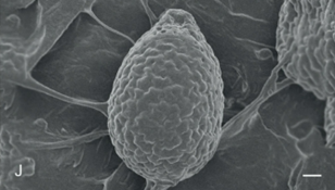
担孢子扫描电镜(SEM)图结果。标尺:2μm
此图片来自文献,如有侵权可联系删除
本词条的创作得到了共同创作者 Uwiling 的帮助。
感谢共同创作者整理物种列表、撰写内容或给出专业性指导!