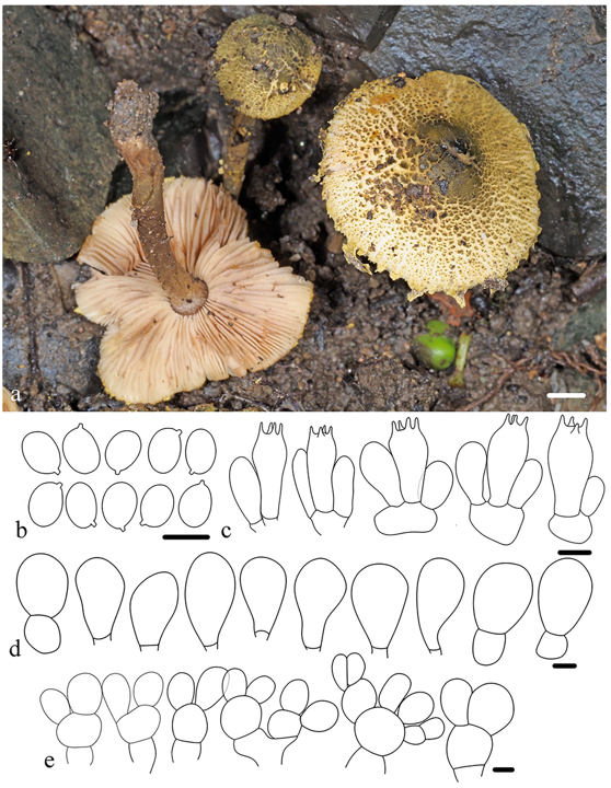
Xanthagaricus luteoviridis R.L. Zhao & J.X. Li, sp. nov. Fig. 14
Chinese name: 黄绿黄蘑菇 (Pinyin: huáng lǜ huáng mó gū)
Fungal Names registration: FN 572558
Etymology: From the Latin ‘luteus’ (yellow) and ‘virens’ (greening), referring to the greenish brown to yellowish brown squamules on the pileus.
Type: China, Jiangsu Province, Nangjing City, Xuanwu District, Zijin Mountain, 24 Sep. 2023, X. Chen, ZRL20236228 (holotype HMAS 282451).
Diagnosis: Xanthagaricus luteoviridis is distinguished by its small basidioma and surface covered with green ish brown, yellowish-brown squamules, which are notably thick and concentrated at the center. The species features an inconspicuous annulus and mostly ellipsoid basidiospores with slightly thickened walls. The cheilocystidia are clavate to broadly clavate with hyaline, thin walls, and the pileus covering is epithelial.
Macroscopic description: Basidioma small. Pileus 15–30 mm in diameter when mature, hemispherical when young, becoming plano-convex to convex with age; surface dry, covered with thick more or less flaky squamules, yellow ish brown to greenish brown, densely and darker at center, becoming lighter towards margin on white background; elsewhere with small scales to fibrillose-squamulose; mar gin with abundant appendiculate, often lacerated velar remnants, concolorous with squamules. Lamellae free, 1–2 mm in width, crowded, depressed around the stipe, broadly ventricose in the middle, pale pinkish to whitish, with 3–4 tiers of lamellulae, with smooth to slightly eroded margin. Stipe 20–30 × 3–4 mm, cylindrical to subcylindrical, surface yellowish brown to brownish, with scattered small scales or fibrils; Annulus not observed. Odor and taste unknown.
Microscopic description: Basidiospores [56/1/1], (3.6)4.1–4.5(4.9) × (2.5)2.8–3.3(3.4) μm, X = 4.3 ± 0.2 × 3.0 ± 0.2 µm, Q = 1.3–1.5, Qm = 1.4 ± 0.1, mostly ellipsoid to broadly ellipsoid, slightly thick walled, smooth, pale yellow in both water and 5% KOH, with a small apiculus, without germ pore. Basidia 13.5–18.2 × 5.1–7.2 μm, clavate, thin-walled, hyaline, 4-spored. Cheilocystidia 13.3–20.6 × 8.5–12.6 µm, abun dant, clavate to broadly clavate, hyaline with thin walls. Squamules on pileus epithelial, composed of agglutinated globose to subglobose, rarely clavate to ellipsoidal cells, 7.9–15.9 × 7.5–12.5 µm, hyaline with thin walls in both water and 5% KOH. Clamp connections absent in all tissues.
Habitat and distribution: Solitary or scattered in soil in broadleaf forest. Currently, known only from one collection from Jiangsu Province, China.
Notes: Phylogenetic analyses based on four loci (ITS, nrLSU, rpb2, and tef1) indicate that X. luteoviridis clusters with X. punjabensis, X. retisporus, and X. lacteus, although the support for this relationship is weak (Fig. 4). X. retispo rus and X. lacteus can be distinguished by their basidioma, which exhibit yellowish to orangish or brownish squamules, lacking the yellowish brown to greenish brown tones seen in X. luteoviridis. Additionally, X. retisporus has larger basidi ospores, measuring 5–6 × 3–4 µm (Yang et al. 2024a). Xan thagaricus lacteus is distinguished by its relatively longer basidiospores (Q = 1.5–1.8, Qm = 1.7 ± 0.1), whereas X. luteoviridis shows shorter and relatively broader basidi ospores (Q = 1.3–1.5, QM = 1.4 ± 0.1). X. punjabensis also has dull yellow orange basidioma with dull reddish-brown squamules. However, it can be clearly distinguished by its significantly larger basidiospores: (4.8) 5.1–5.9 (8.1) × (3.9) 4.1–5.5 (5.8) µm (average 5.5 × 4.9 µm), (Haqnawaz et al. 2023).