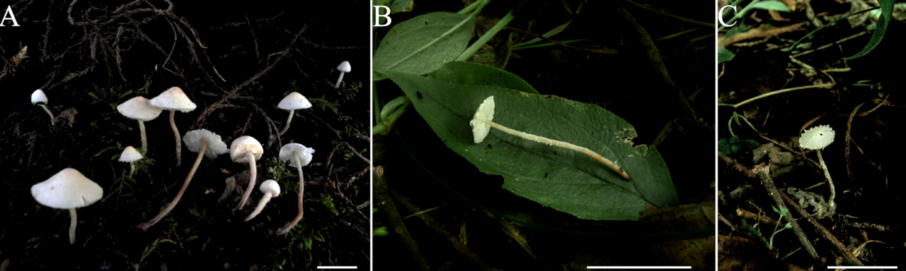
Cystolepiota pseudoseminuda Y.J. Hou, H. Qu & Z.W. Ge, sp. nov. Figure 3A–C, Figure 4C,D and Figure 5.
MycoBank: MB845494.
Etymology: The name refers to the resemblance of the new species to Cystolepiota seminuda [Latin pseudo = false].
Diagnosis: Cystolepiota pseudoseminuda is distinguished from other Cystolepiota species by its slender basidiomata, pulverulent or granulose squamules with a pink to orange tinge that are composed of loosely-arranged inflated cells connected to filamentous hyphae, non-dextrinoid basidiospores with distinct warts on the surface visible under LM and SEM, the absence of cystidia, and the presence of abundant clamp connections. Its ITS, LSU, rpb2, and tef1 sequences differentiate this species from other species.
Type: China, Yunnan Province, Kunming City, Kunming Botanical Garden, on soil under Cupressaceae, alt. 1980 m, 21 October 2015, Z.W. Ge 3795 (Holotype: KUN-HKAS 92275). GenBank: ITS = MN810149, LSU = MN810101, rpb2 = MN820980, tef1 = MN820926.
Description: Pileus 5–18 mm diam, hemispherical to obtusely conical when young, expanding to plano-convex or applanate with a slightly umbonate center with age; surface dry, white to cream, with pulverulent to granulose squamules, fugacious, white, cream, pale orange (5A3), pink (10A3), pinkish-orange to light pink (6A2-5) at the center, turning white to cream towards the margin; margin of the pileus with easily detachable veil remnants. Lamellae up to 2(3) mm broad, free, moderately crowded, unequal, white to light cream, with 1–3 tiers of small lamellulae. Stipe 20–50 × 0.5–2 mm, central, subcylindrical to cylindrical, surface white to cream on the upper portion, with age gradually turning to pale orange (5A3-4), grayish orange (6B5-7), brownish (5D6) to purplish brown (7F7) towards the middle and lower portion with age, with pulverulent to granulose squamules, which are concolorous with those of the pileus; with a fragile and fugacious membranous veil between pileus margin and stipe initially; basal mycelium white. Context thin, whitish. Odorless; taste not recorded.
Basidiospores [100/5/3] (3–)3.5–4.5(–5) × 2–3 (–3.5) μm, Q = (1.21–)1.24–1.85(–2.20), Qm = 1.55 ± 0.19, ovoid to ellipsoid, colorless, thin-walled, inamyloid, non-dextrinoid, metachromatic in cresyl blue; surface slightly punctate-rough under the LM, distinct warts (up to 0.15 µm high) visible under SEM (Figure 4C,D); apiculus small. Basidia 13–23 × 3.5–8.5 μm, clavate, hyaline, 4-spored, rarely 2-spored; sterigmata 1–3 μm long. Lamellar trama regular, made up of cylindrical colorless hyphae, 3–15 μm in diam. Cheilocystidia and pleurocystidia absent. Squamules composed of loosely-arranged globose, subglobose, or ellipsoid, rarely sphaeropedunculate cells, 17–28 × 15–24 µm, smooth-walled, slightly thick-walled, with colorless or sometimes yellowish intracellular pigments; the abovementioned cells are usually attached to hyaline hyphae, 1–6 µm in diam, colorless. Clamp connections present in all tissues (Figure 5).
Habitat and distribution: solitary or scattered on nutrient-rich soil or rotten leaves, distributed in temperate and subtropical zones of southwestern and central China.
Additional specimens examined: China, Gansu Province, Longnan City, Wen County, Chengguan Town, hill near Jiachang Village, on rotting leaves under bushes (Fagaceae, Rosaceae and Coriaria nepalensis), alt. 1700 m, 26 August 2011, X.T. Zhu 574 (KUN-HKAS 73969); Yunnan Province, Kunming City, Kunming Botanical Garden, alt. 1915 m, 9 October 2005, Z.W. Ge 923 (KUN-HKAS 49482).
Notes: Cystolepiota pseudoseminuda is morphologically similar to C. seminuda regarding color and size of the basidiomata. Both species form pulverulent to granulose squamules, non-dextrinoid basidiospores, and clamp connections, while cystidia are lacking. However, the surface of basidiospores of C. seminuda is smooth under LM and SEM, which is in contrast to the warty basidiospores of C. pseudoseminuda. The basidiospores of C. pseudoseminuda are shorter and wider at Q = (1.21–)1.24–1.85(–2.20) and Qm = 1.55 ± 0.19, compared to those of C. seminuda at Q = (1.41–)1.46–2.15(–2.45) and Qm = 1.78 ± 0.22. At least according to the specimens examined in this study, although the characteristics of basidiospores vary strongly between specimens of C. pseudoseminuda and C. seminuda and there is an overlap between the two species in basidiospore size and shape, individuals with high Qm value always belong to C. seminuda and those with low Qm value always belong to C. pseudoseminuda (Figure 6). In addition, there are 92 (out of 722) nucleotide differences between the ITS sequences of the holotype of C. pseudoseminuda and that of the neotype of C. seminuda.
Specimens of C. pseudoseminuda form a robust clade with C. aff. pseudoseminuda 1 and C. aff. pseudoseminuda 2 in in both phylogenies (Figure 1 and Figure 2). The ITS sequences of the three groups are 96% to 97% identical. Their geographical distribution patterns are different: C. pseudoseminuda has so far been collected only in southwestern China and C. aff. pseudoseminuda 1 in Europe and northern China, while all the sequences of C. aff. pseudoseminuda 2 are derived from specimens collected in the Unites States of America (USA). Based on the observation of specimens in this study, no distinct morphological differences were found to distinguish C. pseudoseminuda and C. pseudoseminuda 1 (Figure 4A–D) and no specimens of C. pseudoseminuda 2 were studied morphologically. Further morphological observations need to be conducted on C. aff. pseudoseminuda 1 and C. aff. pseudoseminuda 2.